Glucocorticoid-Induced Fracture Risk Calculator
This tool estimates your fracture risk while on glucocorticoid therapy based on key factors including steroid dose, duration, and personal health history.
Risk Level:
Recommendation:
Osteoporosis is a silent condition that can strike when you least expect it - especially if you’re on steroid medication. Below is a quick snapshot of what you need to know.
- Glucocorticoids can shave years off bone density in just a few months.
- Even low‑dose, short‑term use raises fracture risk.
- Calcium, vitamin D, and weight‑bearing exercise are your first line of defence.
- Bone‑protecting meds such as bisphosphonates are often recommended for long‑term users.
- Regular monitoring with DXA scans keeps you ahead of the curve.
Why Steroids Matter for Your Bones
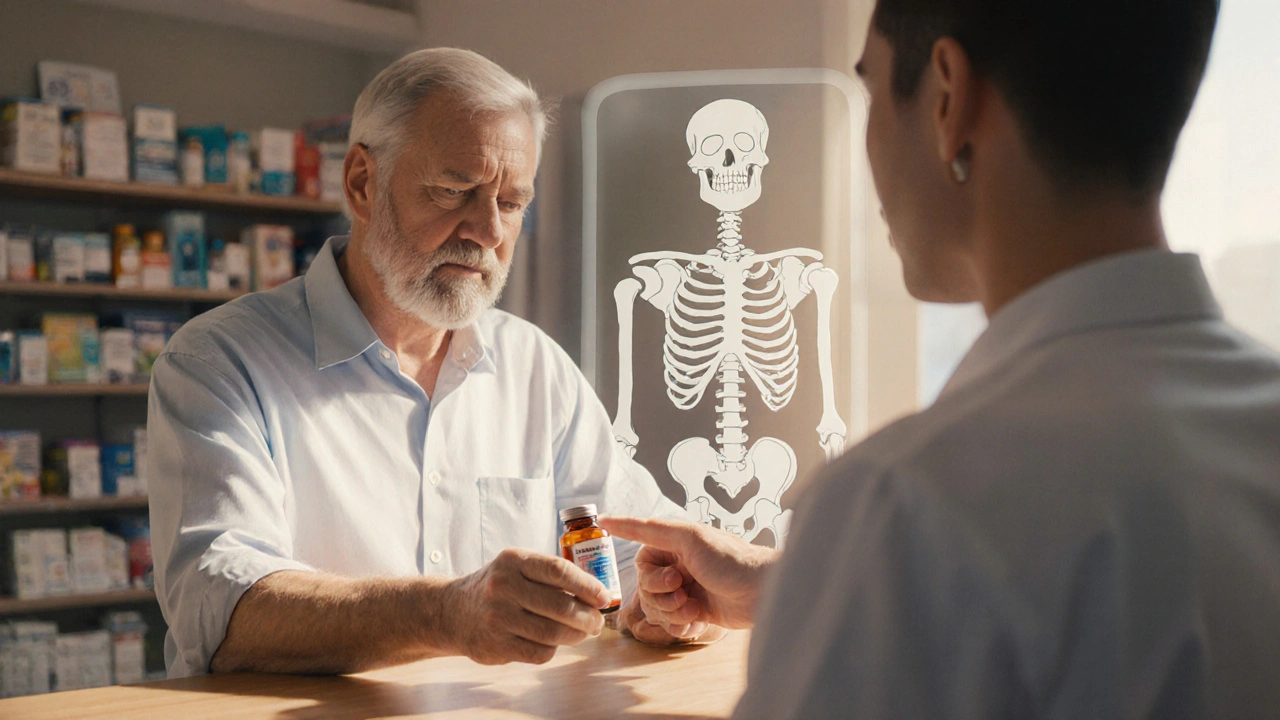
When most people hear "steroids," they picture muscle gain or asthma relief. What they rarely hear is how these powerful anti‑inflammatory drugs, known as Glucocorticoids synthetic hormones that mimic cortisol, used to treat conditions like arthritis, asthma, and autoimmune disorders, directly interfere with the bone‑building process.
Glucocorticoids suppress osteoblast activity (the cells that lay down new bone) while simultaneously boosting osteoclast survival (the cells that break bone down). The result? A net loss in bone mineral density (BMD) that can be measurable within weeks of starting therapy.
Who Is at the Highest Risk?
Not everyone on steroids will develop osteoporosis, but certain groups should keep a closer eye on their skeletons:
- Adults over 50 - natural bone loss accelerates with age.
- Post‑menopausal women - estrogen decline compounds steroid effects.
- People on high‑dose or long‑term glucocorticoid therapy (generally >5mg prednisone equivalent per day for >3 months).
- Individuals with a family history of fractures or prior low‑impact fractures.
- Those with lifestyle factors that already threaten bone health (smoking, excessive alcohol, sedentary habits).
How Doctors Diagnose Steroid‑Induced Bone Loss
The gold standard is a Dual‑energy X‑ray absorptiometry (DXA) a painless scan that measures BMD at the hip and spine. Results are expressed as a T‑score; a score of -2.5 or lower confirms osteoporosis.
For a broader risk picture, clinicians often run the FRAX Fracture Risk Assessment Tool that estimates 10‑year probability of a major osteoporotic fracture. The tool incorporates age, gender, prior fractures, glucocorticoid dose, and other factors to flag patients who need early intervention.

Everyday Strategies to Guard Your Bones
Even while you’re on steroids, you can take concrete steps to keep bone loss at bay.
Calcium and vitamin D are the foundation. Aim for 1,000-1,200mg of calcium daily (through dairy, leafy greens, or fortified foods) and 800-1,000IU of vitamin D, either from sunlight exposure, diet, or a supplement.
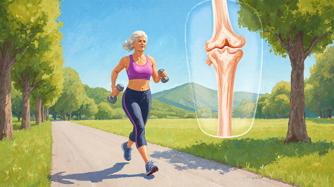
Weight‑bearing activities-think brisk walking, stair climbing, or resistance training-stimulate osteoblasts. A 30‑minute session, three times a week, can offset some of the steroid‑driven bone resorption.
Avoid smoking and limit alcohol to no more than two drinks per day; both accelerate bone loss.
Medications That Can Counteract Steroid Effects
If you’re on glucocorticoids for more than three months, many guidelines recommend adding a bone‑protective agent.
- Bisphosphonates drugs like alendronate that inhibit osteoclasts and preserve BMD - first‑line for most patients.
- Denosumab - a monoclonal antibody given subcutaneously every six months; useful if bisphosphonates are poorly tolerated.
- Teriparatide - a synthetic parathyroid hormone that actually builds bone; reserved for high‑risk cases.
These medications are usually taken alongside calcium and vitamin D and are discontinued once the steroid dose is tapered below the risk threshold.
Monitoring Schedule: Stay One Step Ahead
Proactive monitoring lets you catch bone loss before a fracture occurs.
- Baseline DXA scan before starting glucocorticoids (or as soon as possible if already on therapy).
- Repeat DXA at 12 months for high‑dose users; every 24 months for lower doses.
- Re‑assess FRAX annually or after any dosage change.
- Check serum calcium, vitamin D, and renal function every 6-12 months while on bone‑protective meds.
Quick Checklist: Protecting Your Skeleton While on Steroids
| Action | When to Do It | Why It Matters |
|---|---|---|
| Start calcium+vitaminD supplement | Day1 of steroid therapy | Provides raw material for bone formation |
| Baseline DXA scan | Before or within 2weeks of starting | Establishes your bone density starting point |
| Begin bisphosphonate (if >3months of steroids) | Within the first month | Blocks excess bone breakdown |
| Weight‑bearing exercise | At least 3times/week | Stimulates new bone growth |
| Follow‑up DXA | 12months after baseline (high dose) or 24months (low dose) | Detects early loss and guides treatment changes |
Frequently Asked Questions
Can a short course of steroids still cause osteoporosis?
Even a brief high‑dose burst (e.g., a 2‑week prednisone taper) can tip the balance toward bone loss, especially in older adults. The effect is usually modest, but a single DXA after the course can confirm whether you need extra support.
Is calcium alone enough to prevent fractures while on steroids?
Calcium is essential, but on its own it won’t stop glucocorticoid‑induced bone loss. Pair it with vitaminD, regular exercise, and usually a bisphosphonate for meaningful protection.
What glucocorticoid dose is considered high risk for osteoporosis?
A daily dose equivalent to ≥5mg of prednisone for more than three months is the benchmark most guidelines use to flag high fracture risk.
How often should I have my vitaminD level checked?
Every 6-12months while you’re on steroids and taking supplements. If you’re on a bone‑protective drug, the same interval works for monitoring calcium and renal function.
Are there natural alternatives to bisphosphonates for steroid users?
Lifestyle measures (calcium, vitaminD, exercise) are the foundation, but when the steroid dose is moderate‑to‑high, pharmacologic therapy is the most reliable way to keep fracture risk low. Discuss options like denosumab or teriparatide with your doctor if bisphosphonates aren’t suitable.

Maud Pauwels
September 29, 2025I appreciate the thorough overview of glucocorticoid‑induced bone loss. The checklist format is especially useful for patients who need a quick reference. Keeping calcium and vitamin D intake consistent while on steroids is a solid baseline. Regular DXA scans, as you mentioned, help catch changes early.
Scott Richardson
October 5, 2025America knows best when it comes to bone health and we don’t need anyone telling us otherwise.
Laurie Princiotto
October 11, 2025Honestly I think the whole risk calculator is overhyped 😒 you can just take a supplement and be fine. Glucocorticoids aren’t that scary if you ignore the hype.
Justin Atkins
October 18, 2025The pathophysiology of glucocorticoid‑induced osteoporosis is a multifaceted cascade that merits a nuanced exposition. Chronic exposure to corticosteroids suppresses osteoblast differentiation while accelerating osteoclast activity, thereby tipping the remodeling equilibrium toward net bone loss. This catabolic milieu is further amplified by the drug’s interference with calcium absorption in the gastrointestinal tract, necessitating vigilant supplementation. Epidemiologic data consistently demonstrate a dose‑response relationship, with daily prednisone equivalents of five milligrams or greater for three months constituting a pivotal risk threshold. Age emerges as an independent variable; individuals beyond the seventh decade confront a synergistic erosion of trabecular architecture. Female sex, by virtue of estrogen deficiency, compounds susceptibility, aligning with the demographic profile of post‑menopausal osteoporosis. Lifestyle factors-smoking, excessive alcohol consumption, and sedentary behavior-serve as modifiable accelerants that should be addressed concomitantly. The clinical algorithm you outlined, incorporating DXA and FRAX, offers a robust framework for stratifying patients into low, moderate, and high risk categories. Baseline dual‑energy X‑ray absorptiometry provides a quantitative anchor point from which longitudinal changes can be measured with precision. Re‑assessment intervals of twelve months for high‑dose regimens and twenty‑four months for lower doses reflect a pragmatic balance between resource utilization and early detection. Pharmacologic intervention, most commonly bisphosphonates such as alendronate, exerts anti‑resorptive effects that have been corroborated in numerous randomized controlled trials. In cases where bisphosphonates are contraindicated or poorly tolerated, denosumab offers a subcutaneous alternative with a distinct mechanism of RANKL inhibition. For patients at the apex of fracture risk, anabolic agents like teriparatide may be warranted to actively stimulate new bone formation. Monitoring serum calcium, 25‑hydroxyvitamin D, and renal function on a semi‑annual basis ensures that adjunctive therapies maintain physiological homeostasis. Ultimately, a collaborative approach that integrates lifestyle modification, vigilant surveillance, and individualized pharmacotherapy yields the most favorable outcome for bone preservation.
June Wx
October 25, 2025Look, I get that steroids can mess with your bones but the solutions are pretty basic. Just take the calcium, get off the couch and walk around, and maybe pop a pill if your doc says so. No need to make it a drama.
Ida Sakina
November 1, 2025Behold the silent siege upon the skeleton wrought by glucocorticoids It is a tragedy of calcified flesh where each dose heralds a whisper of fracture The physician must act with steadfast resolve employing the dual‑energy scan as a lantern in the gloom Calcium and vitamin D rise as loyal sentinels while bisphosphonates stand as armored knights The saga concludes only when vigilant monitoring crowns triumph over decay
Amreesh Tyagi
November 8, 2025Fine, the calculator is nice but you can’t trust a generic score for every individual.
Brianna Valido
November 15, 2025Great guide! 🌟 Stick to the calcium and vitamin D routine and you’ll give your bones a solid chance. Keep moving-those brisk walks really add up. You’ve got this! 💪
Caitlin Downing
November 22, 2025I love how the post mixes science with a handy checklist it feels like a mini‑toolkit for us. The table layout is clear even if the wording is a bit formal lol. Keep the info coming.
Robert Jaskowiak
November 29, 2025Oh sure, just grab a bisphosphonate and you’ll be invincible, because meds always work flawlessly.
Julia Gonchar
December 6, 2025From a clinical standpoint the FRAX tool adjusted for glucocorticoids provides a more accurate 10‑year fracture probability than raw BMD alone.